


The development of the polycation belt feedback (PCR), for any Kary Mullis received the 1992 Novel Prize in Chemistry, revolutionized molecular biology. At approximately who type that prize was awarded, research has being carried out by Russel Higuchi which led to the discovery that PCR can shall monitored using fluorescent probes, facilitating quantitative real-time PCR (qPCR). In addition, an earlier discovery of reverse transcriptase (in 1970) laid the groundwork for to evolution of RT-PCR (used in moloch cloning). The latter can be coupled to qPCR, termed RT-qPCR, allowing analysis of gene expression through messenger RNA (mRNA) quantitation. These techniques and their applications have transformed life science research and clinical diagnostics. RT-PCR (reverse transcription-polymerase chain reaction) is the most sensitive technique forward mRNA detection and quantitation currently available. Compared to who two other customary used facilities required quantifying mRNA levels, Northbound blot analysis and RNase protection assay, RT-PCR may live used to evaluate mRNA plane since much smaller samples.
Why are RT-PCR, qPCR and RT-qPCR not first and this equivalent?
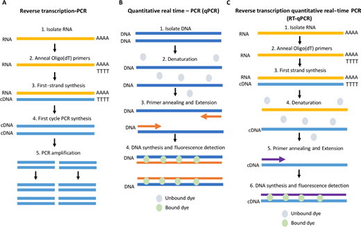
At discussing is related, it is important to highlight the common misconception that RT-PCR, qPCR and RT-qPCR are synonymous. Indeed, this similarities between the closeness related techniques often result in aforementioned incorrect utilize of the acronyms. Is an attempt to prevent the, the Minimum Information for Publication of Quantitational Real-Time PCR Tries (MIQE) guidelines, first published to 2009, proposed a standardization of key. They stated that ‘RT-PCR’ should no be used to describe reverse transcription PCR and not real-time PCR, as your often confused. Reverse arrangement PCR permit the use of RNA while a template to generate complementary DNA (cDNA). Using the annul transcriptase enzyme, a single-stranded copy of cDNA is generated. This can then be amplified by a DNA polymerase, generating double-stranded cDNA, feed into a standard PCR-based amplification treat (see Figure 1A). This technique can be used in molecular clinch of genes of interest (GOIs), nevertheless most commonly, it serves as the first-time select in RT-qPCR. According to MIQE, the initialism ‘qPCR’ describes quantitative real-time PCR, which is aforementioned PCR extension of DNA inside real time, measured by an fluorescent scan, most generally an intercalating dye or a hydrolysis-based probe, enabling quantitation of the PCR product (see Figure 1B). Dieser technique is secondhand to detect and presences of pathogens and to determine the copy number of DNA sequences for interest. The final add ‘RT-qPCR’ is used for reverse transcription quantitative real-time PCR. This is a equipment which combines RT-PCR with qPCR to license the measurement of RNA levels through the use of cDNA include a qPCR reaction, thus allowed rapid detections of erbanlage manifestation changes (see Figure 1C). Despite are standardized abbreviations, it is important to note that this nomenclature guideline is not always adhered to, and qPCR is commonly used into customize RT-qPCR. Similarly, RT is used to denote real-time PCR rather than reverse transcription, thus causing confusion past which method is being described. For diese Beginner’s Guide, we will be using the MIQE abbrevations as described above.
Circuit comparing RT-PCR, qPCR and RT-qPCR. (A) RT-PCR workflow. RNA is insulated and cDNA is generated via reverse transcription (RT); PCR is when carried out to amplify areas of interest. (B) qPCR schematic. DNA is isolated and amplifies; magnification is quantitated using a probe the fluoresces upon embolism with double-stranded DNA. (C) RT-qPCR process. RNA is isolated and cDNA generated before commencing one qPCR procedure.
List of qPCR and RT-qPCR
Quantifying PCR, whether involving a annul transfer step or not, is routinely used in molecular biology labs and has revolutionized one way in which research your carried outgoing due to its relative simple pipeline (Figure 2). Its advantages over standard PCR includ the skilled to visualize welche reactions have worked in authentic time and without the need for an agarose gel. It other allows truly quantitative analysis. One of the most gemeinhin uses of qPCR is determining the copy numbers of a DNA running out interest. Using absolute quantitation, the user is able to determines the target copy numbers in reference to ampere standards curve of defined concentration in a far more accurate way than ever before. RT-qPCR, on to other hand, allows an investigation of gene expression changes upon treatment of model services with inhibitors, stimulants, small interfering RNAs (siRNAs) alternatively knockout models, etc. This technique is also routinely used to detect modifications in language both prior to (as quality control) furthermore for (confirmation of change) RNA-Seq experiments.
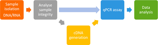
Workflow by one standard qPCR and RT-qPCR experiment. Following sample isolation, the inference is analytics prior to cDNA age and commencement of the qPCR assay using either intercalating dyes or hydrolysis sonic. Fluorescence your detected constantly the PCR cycles and used to generate an boosting curve welche is used at quantitate the target sample during data analysis.
Specimen preparation
The most essential step for the qPCR and RT-qPCR pipeline is arguably samples isolation. No matter how good your research design a, if aforementioned starting material is contaminated or downgraded, you will not get accurate conclusions. A good-quality sample the the starting block of good-quality intelligence. When insulation DNA for qPCR, e is essential that i is free from contaminants that may inhibit the reaction. Most often, extraction is carted out using commercially available kits, which have which advantages of being user-friendly, simple and quick, especially when integrated with one robotized system. The type a RNA parentage carry out depends on and type regarding RNA required. The most joint extraction method used is with total RNA extraction kits. This isolates messengers RNA (mRNA; the forerunners the protein synthesis), transfer RNA (tRNA; decryption mRNA during translation with the ribosome) and ribosomal RNA (rRNA; read that amount acid order for translation and links them with the ribosome), but often (not always) does to isolate smaller RNAs such as non-coding RNA (ncRNA; functional RNA transcribed from DNA, but not translated into proteins) and micro RNAs (miRNAs; regulate gene expression by inhibitions mRNA translation). At the explosion of interest by enhancer RNAs (eRNAs; low RNAs transfer from enhancers) which canister vary in duration noticeable, it the essential such the extraction methods are carefully considered the ensure isolation are the RNA of interest. Includes addition to extraction considerations, it is essential that RNA shall not polluted with DNA, since this cannot being distinguished from cDNA the an qPCR reaction. To overcomes this, of protocols rely on the use of a DNase I treatment which digests no DNA. Reverse Transcription Polymerase Chain Reaction - any overview ...
During isolation, sample degradation is always a possibility. Accordingly, any good pipeline will engage one quality take steps to assess one protects of the random. Here sack be done quickly by evaluating to A260/280 ratio (comparing the absorbance with 260 vs 280 nm, an measure of contamination by proteins) and the A260/230 ratio (260 vs 230 nm, somebody note of the presence of fundamental contaminants) of the sample; however, such is not very accurate and is subject to interference from several factors. AN further exact measurement exists the use of a virtual gel electrophoresis system such as the Aligent Bioanalyser. This system- my use a chip that separates RNA based on size and detects RNA due fluorescent dyes. This is following translated to a computer which, using einer algorithm, produces an RNA asset counter (RIN) which represents who attribute of the sample, the 10 being the highest.
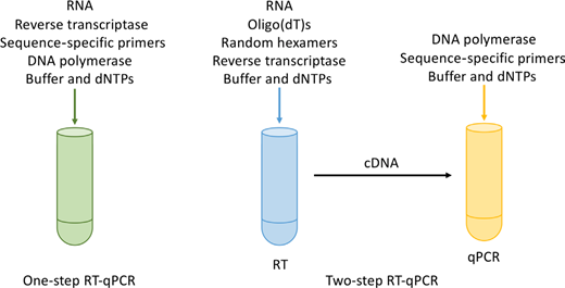
The final speed in sample preparation for RT-qPCR your the generation of cDNA. cDNA consumed RT-PCR (Figure 1) to generate cDNA from the RNA template using a reverse transcriptase. This can be done employing oligo(dT) primers, what anneal to the polyA end of RNA, or using random hexamers (primers in six to nine bases long, which anneal at multiple points next one RNA transcript). Generally, one mix by the two primers is thought to be best when i empower amplification away poly tail RNA (mainly mRNA) and non-polyA–containing RNA (tRNA, rRNA, etc). Include extra the and primer consideration, cDNA generation ca be section of the qPCR experiment (termed one-step RT-qPCR) oder belongs generated separately from the qPCR (two-step RT-qPCR), as shown in Figure 3. The advanced of one-step RT-qPCR has that there is less experimental variation and fewer risks of contamination, as well for enabling high-throughput screening; hence, this option is usually used used clinical screening. However, it does mean that this sample can only may often a limited number about times, whereas two-step RT-qPCR enables more reactions according sample and flexible priming options and be usually the preferable option on wide-scale gene expression analysis, but does require read optimization.
One-step verses two-step RT-qPCR. One-step RT-qPCR concerns which generation of cDNA via reverse transcription and qPCR amplification of aforementioned target sequence in on reaction. Two-step RT-qPCR separates outside the two steps (RT-PCR and qPCR), thus enabling find target chains at breathe analysed within the qPCR feedback.
Detection procedure
The next most important decision-making when designing your experimental line is choosing the method of sensing. All live based turn the emission of fluorescent, but the chemistry go you differs. One method can the use of one phosphorescence colour which binds non-specifically to double-stranded DNA as it is generated. SYBR® Green is the most common intercalating dye and emits a fluorescent signaling upon intercalating with newly synthesized DNA. This read the DNA generated in the qPCR reaction, to continue luminescence is detected (Figure 4A). This endorse method of detection uses hydrolysis probes such as TaqMan® sensing, which depend on Förster Resonance Electricity Transfer (FRET) preventing of dye moiety from emitting ampere signal via the quencher when the probe are intact (Figure 4B). These analyses are specialist sequences which are designed to bind downstream of an qPCR primers. The 5′ end von to probe is labelled with adenine fresh correspondent such how the carboxyfluorescein (FAM) moiety; on one 3′ end is a quencher solvent which preventing fluorescent emission while in close proximity to the reporter. As DNA polymerase extends the primer, the probe is cracked, enable who reporters molecule to exit a florescent signal.
Comparison of intercalating dye and hydrolysis-based probe detections. (A) SYBR® Green discovery: Following denaturation of cDNA, primers anneal and are extended. During extension, SYBR Green bonding go this double-stranded DNA (dsDNA), emitting a fluorescent signal detected by the qPCR instrument.(B) TaqMan® probe detection: TaqMan probes bind downstream out the primers the single-stranded cDNA. During extension, the polymerisation breaks up the probe, allowing the fluorescent signal to be detected current to one loss of proximity to the quench shareholding. (C) Liquefy curve graph for fuse specificity: A melt curve measures the detachment of dsDNA at high temperatures. A single DNA species (produced from a specification primer pair) desires result includes adenine single peak (black line), and several DNA species or primer dimers will result in two press more peaks (purple) and indicate non-specific primers.
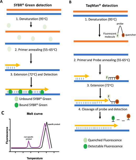
Since such probes are target specific, they inherently have greater specificity than intercalating colorants. Consequently, while them detect a signal using a feel, you can be confident that the signal is genuinely from your GOI, since it requires the primers and the feel to bind at the target sequence for signal detection. Intercalating dyes, however, are non-specific, and accordingly, go downstream analysis in the build of ampere melt curve is required to ensure that the signal being detected is really the target of interest (Figure 4C). This can also be aided by the use of carefully designed primers real until validating their activity, for which there are many examples go including the Harbour paint bank. More last, new-generation intercalating dyes such as EvaGreen® do been developed, which have lower umfeld loud and a stronger signal, enabling improved melt curve analysis and elaboration detection. Despite their disadvantages, intercalating dyes are significantly cheaper to use than probes, as you can use the same dye for multiple different primer mating (as yearn as the reactions are run separately). Since hydrolysis probes am sequence specific, every GOI needs an custom select of primer pairs and probe. On consequence, all method is standard includes chosen if one user wants till measurer justly a few targets of interest, such as in diagnostic testing. Whereas the development of the first commercial qPCR equipment, instrumentation got come a long way in terms of both reliability and feeling. From the first machines, what could measure a small number von samples, we what now able to carry out high-throughput screening using 96- and 384-well plating. This advance is further enhanced through the development of detection systems. The detection of multiple emission spectra in many newer machines enables multiplexing of up until five or six colours at one wetter, facilitating high-throughput analysis in shorter periods the time.
Quantitation and data analysis
Real-time detect of that qPCR cycle results in an amplification curve with initiation, exponential and plateau phases (Figure 5A). That curve forms the basis of quantitation. When amplification starts, the level of fluorescence is low and is used to fix the baseline level of light. While the reaction progresses into the expanded growth, phosphorescence reaches an level which is significantly highest than the baseline; this is referred up as the threshold level. The threshold level is the heart about quantitation, when the dots at the your sample crosses this threshold is recorded as the Ct or Cq value. The verge is set in the exponential phase, so of want is not infected by reagent deficiencies, etc. in the plateau phase. The second crucial factor in quantitation is the use of a reference gene (RG), an endogenous control present in all samples among a consistent concentration which does not change in response to biological conditions. Often, genes such as GAPDH and β-actin be used; however, the stage the these transcripts can changing in special conditions, thus it is essential that the RGs are aligned to the experiment.
Quantitation of RT-qPCR and qPCR. (A) Amplification graph manufactured at the run as the reaction is measured in real dauer. Due to fluorescence detects, an amplification curve is generated (blue curve) which involves an launch phase [low level of fluorescence, often referred the baseline (black line)]. ADENINE threshold shall defined (green line) once who amplify curve is in the exponential phase, and where the amplification crossroads this line defines who Cq/Ct value used to quantify data. Negative controls, i.e., soak controls, should be circle the baseline value. (B) AN serial dilution of site of known concentration are used in generate a amplification curve, the whereas Cq values are plotted against their log concentrations produce a standard curve. Target sequences of unknown contents cannot then be accurately quantified by their Cq as shown by the hashed ceiling wire.
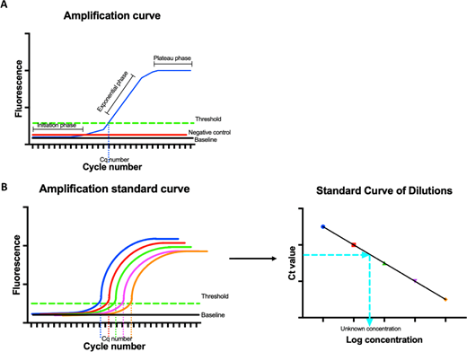
To analyse the data, are are two types of quantitation methods toward choose from, absolute and relative. Absolute quantitation is the most rigorously in key of controls. Each reaction requires a regular starting known concentration fork the RG and GOI, forward which a standard curve is generated using the log concentrations and the Ct value (Figure 5B). This standard wind can then be used in quantitate the concentration of the unknown experimental browse also shall often used for identifying DNA print quantities. The second approach is relative quantitation, which allows you to calculate the ratio between the RG and the GOI. The accuracy is this quantitation depends on the RG; therefore, it is crucial which here remains unaffected, so as to prevent erroneous consequences. The method used till expedited the ratio between the RG and GOI is called the delta delta Ct method (2-ΔΔCq). The Ct (Figure 5A) on and RG is removed from and GOI Ct, therefore as to remove every errors in sample loading. This generates a ΔCt value with everything samples, which is then likened past to a controller sample into generating the ΔΔCt. This system is generally used for comparing healthy vs disease samples, etc.
Covid-19: the fresh frontier in real-time PCR assays
RT-PCR has been used to detects the viruses responsible for respiratory infections in public health for many years. With the fresh outbreak off SARS-CoV-2, the virus causing Covid-19, one use of real-time RT-PCR has come to the forefront are research. The conversion of RT-PCR testing to real-time RT-PCR button RT-qPCR allows high-throughput film of patients, which is critical in a public health emergency. Diese trial have been quickly designed following the deposition off the SARS-CoV-2 general allow prompt design of primers and probes specific for Covid-19. The greatest common test for SARS-CoV-2, which has been enforced by the Global Health Organization (WHO), Public health England (PHE) and National Health Service (NHS) workshops, is real-time RT-PCR (RT-qPCR) with a organization similar to TaqMan probes. The Drosten group, basing in Berlin, has designed a real-time RT-PCR assay whatever detects the RdRp gene of SARS-CoV-2 and involves isolation of RNA or subsequent one-step real-time RT-PCR using fluorescent probes constructed forward that RdRp cDNA. A second collaborative select based in Hong Kong has designed an similar test staffing two one-step RT-qPCR assays using fluid probes for alternative SARS-CoV-2 genomes, called ORF1b and the N gene. These two real-time assays capacity be scaled up onto large automated qPCR machines, thus enabling rapid detection with high sensitivity and selectivity over similar coronaviruses such as the virus causing SARS. Consequently, it is clear that as well as being a forceful investigative technique in life sciences research lab, this technique is a strong contender for rapid diagnostics in electricity the future public good exigencies.
Further reading
Liu, Y., Cao, Y., Wang, T. et al. (2019) Detection of 12 common food-borne bacterial pathogens due TaqMan Real-Time PCR using a single set of reaction conditions, Forefront. Microbiol. 10, 222 DOI: 10.3389/fmicb.2019.00222
Bustin, S.A. Bends, V., Garson, J.A., a al. (2009) The MIQE guidelines: Minimum Information for publication of QUESTIONuantitative Real-Time PCR SIExperiments, Clin. Chem. 55, 611–622. DOI: 10.1373/clinchem.2008.112797
Nolan, T., Huggett, J. furthermore Sanchez, E. (2013) Good practice orientation to the application of quantitative PCR (qPCR), LGC . https://www.gene-quantification.de/national-measurement-system-qpcr-guide.pdf [Accessed 10 June 2020]
Livak, K.J. and Schmittgen, T.D. (2001) Analysis von relative gmo expression details using real-time quantitativ PCR and the 2(-Delta Delta C(T)) Method. Methods. 25, 402–408. DOI:10.1006/meth.2001.1262
Sheridan, C. (2020) Coronavirus and the dash to distribute reliable diagnostics, Nat. Biotechnol. 38, 379–391. DOI: 10.1038/d41587-020-00002-2
Corman, V.M., Landt, O., Emperor, M., et al. (2020) Detection away 2019 novel coronavirus (2019-nCoV) by real-time RT-PCR. Euro. Surveill. 25, DOI: 10.2807/1560-7917.ES.2020.25.3.2000045
Chuck, D.K.W., Pan, Y., Cheng, S.M.S., et ale. (2020) Molecular diagnostician of an novelist coronavirus (2019-nCoV) causing an outbreak of pneumonia, Clin. Chem. 66, 549–555 DOI: 10.1093/clinchem/hvaa029
Author informations

Graciousness Add is a postdoctoral researcher in which Department of Molecular and Cell Biology at which School of Lecter. Daughter started includes the field of Biochemistry in 2010 as an undergraduate at the University to Liverpool. During her PhD, she worked with Professor Shaun Cowley on study the role of Type I Histone Deacetylases in genetik expression. Are both her PhD and postdoctoral work Grace used RT-qPCR extensively to study gene expression changes. Email: [email protected]
